Fix or Replace That Digital X-Ray?

By Bryan Delano

The film X-ray era is fully behind us, and we are now entering the second and third generations of digital 2D panoramic/cephalometric technology. The first models of digital X-rays, available in the late 1990s, were film-to-digital upgrades. In the early 2000s, direct digital 2D options were introduced. Today, practices have the option of implementing either new or pre-owned digital X-ray technology at a fraction of the cost of just ten years ago.

As some digital panoramic models reach almost twenty years of age, and most first-generation cone beam units are over ten years of age, warranties have expired, repairs are more frequent, and the cost and availability of parts is challenging. Additionally, software drivers that control these units are not keeping up with modern computer operating systems, limiting available compatible options in the event of a PC failure. When faced with these obstacles, a practitioner has the option to either invest in a costly repair or purchase a replacement unit.

Just like any modern technology, repairing an X-ray unit can range from a ten-dollar simple fuse replacement, to tens of thousands of dollars when replacing a failed sensor. Please see the guidelines below when faced with X-ray component failures.

Digital sensor failure

Unfortunately, there are few, if any viable sensor repair resources, so the replacement of the actual sensor is often the only option with a digital sensor failure. Some X-ray models such as Sirona, Planmeca and Instrumentarium are formatted with cartridge-type sensors that can be moved between pan and ceph. A pan sensor can only be used to capture a panoramic image, but a cephalometric sensor can be used to take a pan OR a ceph. Switching the sensor between the pan and ceph is easy and can provide a long-term solution or can buy time to research other options. It’s never good to feel pressured to make an expensive purchase quickly, so this option can help extend the decision-making period. Purchasing a new sensor can range from $3,000 to $15,000, depending on the manufacturer, but there are other options to consider. For example, often you can find X-ray sensors on eBay that offer Buyers Protection. This allows a buyer to receive the sensor, test it thoroughly, and return it for a full refund if it is defective.

We don’t know what is wrong

Many dealer technicians rely on the X-ray manufacturer for support. When your tech arrives, they inevitably will immediately contact the manufacturer for help. The older generation machines are often misdiagnosed at this point, and the tech will suggest ordering or trying up to several parts, which can be very costly. Some of these parts may be needed and others will not. The challenge of only ordering one part at a time could result in delayed repair time with multiple shipments and on-site tech labor charges. When ordering several parts at a time, however, make sure to ask if the unused parts be returned for full credit or a refund with a re-stocking fee, or if they cannot be returned at all. Major replacement parts can cost anywhere from $3,000- $10,000 (not including service), even if the sensor is not involved. Warning: Buying parts other than sensors on eBay can be limited or difficult, and often dealer technicians will refuse to replace these parts due to liability issues.

I’m sorry, this unit is discontinued, and parts are no longer available

By law, X-ray manufacturers must make parts and support available for their equipment for around eight years after the machine was last sold. For example, if your machine was manufactured in 2007 and sales stopped in 2010, they are obligated until 2018 to provide replacement parts and support. That doesn’t mean if your machine is dated 2007, then you are already out of luck. If that model is currently still manufactured, then you have a long runway for parts availability. Many manufacturers are still providing parts and support beyond the eight required years, but the challenge for them is that these parts were not made by their own company, but by third-party suppliers. If those suppliers choose not to continue manufacturing the desired part past the eight-year requirement, then the X-ray manufacturer is left with only the parts that they have on their inventory shelves. This is a common issue with many X-ray manufacturers.

Replacing the unit

Replacing an X-ray unit is a costly proposition. Fortunately, digital panoramic unit pricing has come down significantly in recent years. In addition, many quality pre-owned X-ray options are also available. The challenge now becomes that your X-ray is down and you need a replacement in a short period of time. This time factor could limit your options and ability to negotiate the best price. Many X-ray manufacturers have ended exclusive distribution deals, so you can shop for the same X-ray model from several distributors for the best pricing. Depending on the repair status of your current unit, you may be able to receive some trade-in value based on the remaining parts. Perhaps this is the practice’s impetus to choose and upgrade from 2D to 3D. If you want to “buy” some time for additional research, you can also ask the new / used X-ray vendor to fix your unit with borrowed / loaned parts until the new X-ray is purchased.

So, when faced with the challenge of repairing or replacing your X-ray, ask yourself the following questions:

  • What are the costs of the repair? Do they exceed the costs of purchasing a new or pre-owned unit?
  • Can I get away with “patches” such as swapping a sensor between a pan/ceph unit or buying parts on eBay?
  • If my X-ray is over eight years old, are parts still available?
  • Can I allow enough time to research my options and compare pricing between vendors?
  • Is now a good time to consider upgrading from 2D to 3D?

When possible, the best practice is start planning ahead for older X-ray equipment replacement. But, since you cannot always predict equipment failures, it never hurts to start researching your options today.


Sharing Cone-Beam CT Images Online

By Dr. Dan Grauer

When diagnosing and treatment planning interdisciplinary patients, have you ever sent your three-dimensional images to a colleague? Have any of your patients requested a copy of their records for a second opinion? Or maybe, a patient declines a radiograph because another orthodontist has recently taken a CBCT image of the patient? In all of these instances, you will need to communicate with the other office to initiate the transfer of CBCT images. The purpose of this blog is to describe different methods used to share patients’ CBCT records via online means.

Images acquired in your office are requested by a second orthodontist/dentist:

The first question that will need to be answered is whether the other office has the possibility of viewing and analyzing the images in three-dimensions. In a few instances, I have found myself trying to transfer a full three-dimensional file, when the second orthodontist just wanted a cephalogram and a panoramic radiograph. If this is the case, your software will probably allow you to create a synthetic cephalogram and panoramic radiograph that can be emailed through a HIPAA-compliant email account. If the second orthodontist requires a three-dimensional image, two case scenarios are possible:

Case scenario 1: Second orthodontist owns software to read and visualize CBCT images.

In this case, your software is able to export the CBCT Images in DICOM format (Digital Imaging and Communication in Medicine). DICOM files are large, and a file transfer application is needed. Once transferred, these can be imported into the software of the second orthodontist for visualization and analysis.

Case scenario 2: Second orthodontist does not own three-dimensional imaging software.

Under this case scenario, the second orthodontist would need both the CBCT images and a three-dimensional viewer. Three main options are available.

Option 1: If you own a CBCT machine, your software is generally able to create a file that includes both the image data and a basic viewer. The files created are large and can be transferred with a file transfer application.

Option 2: Anatomage offers the possibility of uploading your CBCT images to the cloud, and these can be accessed online through Anatomage’s application, which acts as a visualization tool. At this point the software is in Beta-version and can be accessed at www.anatomagcloud.com. You, as the generating office, will need to upload the images to the AnatomageCloud database and use this application to allow the second office to access the specific patient images. The access is granted with a link embedded in an email. After receiving authorization to access the images, the second office will be able to access the images online without the need of downloading them or installing any software.

Option 3: Dolphin Imaging software offers a complimentary viewer, https://www.dolphinusers.com/dolphin-imaging-viewer/. The receiving doctor can view 3D images by downloading and installing the Dolphin Imaging Viewer software. Files are transferred in DAZ file format. This file format is proprietary to Dolphin Imaging, and the files are created by the originating doctor through Dolphin Imaging 3D Software. This option 3 would work also in Case Scenario 1, when both doctors use Dolphin Imaging 3D software, but it is important to note that only the unprocessed images need to be transferred, such as the DICOM file; the viewer is part of the software downloaded by the receiving office.

Images acquired by other offices:

Images that you receive from other offices should be requested in DICOM format. This will permit you to be able to import these into your 3D software. If you obtain the file in a different format than DICOM (that often includes the viewer), the analysis and measurement possibilities are limited; this is because your 3D software most likely includes all the features that you may need while visualizing and measuring 3D Images. If both offices use Dolphin Imaging 3D Software, a proprietary format DAZ can be used to transfer and share images. The advantage of this approach is that all patient images, including both 3D and 2D images, are shared simultaneously.

In summary, with Cone Beam CT becoming more popular in practices, sharing 3D images with other treating doctors or practices requires some additional steps. The first step is to initiate the conversation with the second office to establish the best system to use to share images. The advantages of 3D images over traditional 2D images are beyond the scope of this blog, but once you become accustomed to a transfer and visualization system, the collaboration between doctors and patient care may improve.

CBCT Imaging for your Practice: Is Now the Time?

By Dr. Kenneth R  Webb

As I walked the exhibit floor at the AAO Annual Session in San Diego this past April, I stopped and talked to several of the company reps displaying the latest generation of CBCT scanners. Competition is good – what struck me is how far this technology has come in such a short period of time. Effective dose, image quality, and versatility are three areas where the advances are most impressive. I have witnessed these advancements first hand in my practice where we have been using CBCT since 2012 and recently upgraded a satellite office from a 2D digital Pan / Ceph to an “Ultra Low Dose” current generation CBCT scanner. I’d like to share some of my thoughts about the 2D to 3D transition.

Effective Dose
I encourage anyone interested in CBCT technology to search for studies authored by Dr. John Ludlow and his team. Dosimetry data and image quality for many CBCT scanners (both older and current generations) have been researched in great detail. Comparative data for 2D dental radiographic imaging (both intra and extra oral) is also available. By replacing our 2D Digital Pan / Ceph with a current generation CBCT scanner we have reduced the effective dose of our diagnostic records imaging by approximately 60%. (Ludlow JB, Walker C. AJO-DO, 2013;144 (6): 802-817) This is accomplished with one, approximately 5 second, ultra-low dose CBCT scan compared to the two longer duration exposures (pan then ceph) required with 2D imaging. Certainly a win-win for our patients.

Versatility
What else can we use the 3D data for? Digital models: used to assess the inter-arch and intra-arch relationship as well as aiding the determination of arch length and arch width requirements and assessing arch symmetry. Modelling labs can produce articulated 3D study models from the CBCT data (DICOM) files.

STL files of the patient’s dentition and occlusion can be uploaded into whatever software you may be using in your office for 3D model storage and viewing. Additionally, these models can be used (with appropriate software) to produce treatment simulations by “moving” individual teeth or the arches as a whole. Imagine completing your diagnostic records with Intra / Extra Oral photos and one 5-second CBCT scan!

Viewing the Diagnostic Data
The software that comes with a CBCT scanner can easily produce the customary 2D diagnostic images that we are used to evaluating (panoramic, lateral and A-P ceph). In addition – you see everything! The data can be viewed in sagittal, axial or coronal sections (slices) or as a 3D “volume rendering” which can be rotated and sliced (“clipped”) at will. So why is this additional information gained from CBCT imaging important?

Pathology
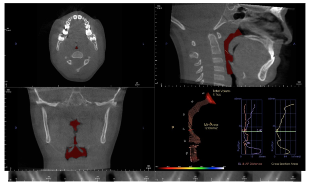
You will see pathology in the 3D data that isn’t visible with standard 2D imaging. When pathology is visible in 2D, the 3D data can more accurately ascertain location, extent, and character of the area of concern. This is beneficial to our patients.

Developmental Anomalies
One of my first revelations when we began imaging in 3D was the prevalence and extent of “individual anatomic variation”. But how much is too much? We have made referrals to medical specialists for significant developmental anomalies in the cervical spine, nasal cavity and paranasal sinuses. Patients / parents are appreciative of the thoroughness of our diagnostic process that includes 3D imaging.

Asymmetries
They get their own paragraph! Your patient smiles at you and you notice a vertical asymmetry and occlusal plane “smile” cant. Is it caused by hemimandibular hyperplasia, condylar hyperplasia, a unilateral expansive lesion in the maxilla, a growth response to unilateral progressive condylar resorption (to name a few) or a growth response to a foreign object lodged in the nasal cavity? I’ve seen them all. Similar clinical presentations – different treatment plans.

TMJ
Condylar position in centric occlusion, condylar size and shape, glenoid fossa morphology, condylar osseous morphology, joint space, findings suggestive of degenerative joint disease – both active and stable. It is hard to argue that these are not important considerations in our diagnostic process.

Airway
The value of volumetric and cross-sectional analysis of the naso – pharyngeal airway and its relationship to orthodontic diagnosis and treatment planning has, and continues to be, studied in great detail. Is the growth and development of a patient who presents with narrow arches, an anterior open bite or anterior crossbite, tongue thrust and a strong mouth breathing pattern secondary to adenoid / tonsillar hyperplasia, a deviated nasal septum, environmental allergies or restrictive airway dimensions in general? Should your imaging modality include an assessment of these areas?

And there is more…
Orthognathic surgical treatment planning, TAD placement guidance, precise localization of ectopic and supernumary teeth, and assessment of treatment progress – including evaluation of root torque. All are possible or enhanced with 3D imaging. And by managing scan parameters (field of view, scan time and voxel size) at an effective dose equal to or less than 2D imaging modalities.

So, if you haven’t brought this technology into your practice – is now the time?

At the 2017 AAO Winter Conference in Ft. Lauderdale, Mr. Chris Bentson reported on a survey of recent (2016) U.S. Orthodontic Residency graduates: 88% responded that they had used CBCT imaging for diagnosis and treatment planning during their residencies including 21% that used it on all patients.

For the 2013 graduates, the responses were 76% and 9% for the same questions.

Based on these statistics, are we that far away from 3D imaging being included in the “standard of care” discussion? Embracing new technology is not easy but the value added for the practitioner and our patients is significant. The orthodontic educators who expertly taught my generation faced a similar situation during their careers with a then relatively new technology: panoramic radiographic imaging. My generation faced the challenges of transitioning from analog imaging (film) to digital.

Change is not easy. The transition from 2D to 3D imaging in your practice will require a financial and educational commitment. The whole staff will be a part of this paradigm shift. A positive – 3D imaging will be a differentiator for your practice. If you are seeking an associate, partner or complete transition, 3D imaging will make your practice more attractive to the next generation of orthodontists.

3D imaging will benefit you and your patients. Is now the time?

What Your Email Address Says About You

By Steve McEvoy, Technology Consultant

Sending business emails ending with @Netcom.com, Aol.com, Earthlink.com and soon Yahoo.com are tell-tale signs you aren’t keeping up. People notice and may ask you if you are still driving the ‘72 Ford Pinto as well.

Using personal accounts from your Internet Service Providers (ISP) also looks old school like Comcast.net, TimeWarner.com, RoadRunner.com, SBCGlobal.net.

There is no rule making us keep up, or against driving a ‘72 Pinto, but I’d argue if you are in a marketing battle for new patients, this is just one small aspect you can easily improve.

For work, you really should have an email for your practice’s domain.   [email protected] or [email protected], for example. People expect this, and when they see [email protected], you send a subtle message that isn’t positive.

You might certainly have a private, personal email account as well, but even for that, you should consider using something like [email protected] or [email protected].

This should also apply to any of your staff using email on behalf of the practice.

There may be other motivating reasons as well. Companies offering email services for free (AOL and YAHOO) that aren’t thriving financially in their core business won’t be putting development resources into keeping products current, safe and secure.  The news is full of stories about hacked email accounts and ransomware which typically makes its way into your computer via email. Companies like Google, Microsoft, and Apple have reputations to maintain and will have the resources to keep up.  If you are going to use a free email service, I might suggest you look to one of the major players.

Changing your primary email account is always a major hassle, and this is likely what has kept you from making the switch.  Setting up a new email account, and merely forwarding all the email from your old account indefinitely isn’t the right solution. Your old email account can still be hacked, and the company can still go out of business.

The steps generally are:

  • Setup your new email account – and take this opportunity to make sure the password is a hard one. I’d suggest you setup two-factor authentication with it as well.
  • Link your new account to your PC, phones, tablet, etc.
  • Using your old email, notify EVERYONE in your contact list that you are changing your email to the new one effective immediately.  You can do this in one mass email, but be smart about it and put all the recipients in the BCC list so they don’t see everyone else you emailed the list to. Make yourself the only official To:
  • Configure your old email account to forward to your new one (for a while).
  • After a month, check your old account to see if anyone you know is continuing to use the old account, then contact them directly to start using the new address.
  • After another month, turn off the forwarding from your old account and delete it at the vendors. Gone forever, but no risk of hacking.

You can also always enlist some help from your IT person, they should be well familiar with the process.

Creating Videos for Your Orthodontic Practice

Humans are visual creatures. Simply put, we process visual data better. Research shows that more than 80 percent of human learning is done visually and that humans process visuals 60,000 times faster than text.1  As such, creating compelling, easily sharable videos is one of the best ways you can choose to promote and showcase your orthodontic practice. Thankfully, creating low cost, high impact videos online has never been easier. With just a small amount of effort and know-how, you can make a lasting, positive impression on potential patients and set your practice apart from competition in your area.

Videos that introduce yourself and your staff, feature patient testimonials and showcase your practice and the services you offer is a key way to build trust and let potential patients get to know you before actually ever stepping foot in your office. As a bonus, videos hosted on your practice website can also improve how your site appears in search engine results…meaning increased web traffic.

Videos can easily be created without a major investment in equipment or software.  Although they do take some planning and time, especially early on, the investment is well worth the effort. But, if you are trying to keep your costs low, a newer model iPhone or Android device you may currently have in your pocket offers outstanding video capabilities. (Just be sure you’re shooting in horizontal vs. vertical mode.)

Regardless of what equipment you use, following these basics.

  • Shooting a video can be intimidating at first. Just always try to focus on sound and picture quality.
  • Shoot your video in 720p or 1080p resolution.
  • Pre-plan your video. Have a goal in mind. Create and work from an outline so you don’t go off-track.
  • Be sure to add captions to your video. This is because up to 85 percent of videos watched and shared socially are viewed without.  Captions are added via a video editor’s text feature.
  • Make the first few seconds of your video visually impactful. Your goal is to capture your viewer’s attention quickly, within the first 3 seconds.
  • Give your video a compelling title or headline.
  • Brevity is key. Be efficient with words and visuals.

As you begin planning your videos, consider:

  • Stability is key. Invest in a small tripod, monopod or desk stand – your videos will be of much higher quality if you can prevent excess camera movement.
  • If you are producing a video for patient education, you may want to invest in a high quality consumer camcorder with an external microphone.
  • While professional lighting is always preferred for marketing and practice website videos, natural lighting is usually sufficient. Just film in a brightly lit room and make sure there is enough light on your subject’s face.
  • Make sure to have your interviewee or one of their parents sign a photo/video release form giving you permission to use the video you just shot. These forms are available at aaoinfo.org. Log on; then go to Practice Management > Forms and Releases. Scroll down to Practice Management Forms & Materials; then click on Photo Releases.
  • Think outside the box. Videos can be an extremely powerful communication tool for patient education and staff training.

Once you’ve captured your video, you’ll want to begin editing. Editing video means trimming and arranging your video. Video editing software is also how you can add transitions, music, captions and titles…as well as export the movie file that you’ll host on your website or social media channel. Thankfully, there are easy-to-use and affordable options available that will allow you to quickly transform your raw footage into a finalized video for your web page or social media channels.

For users on the Mac, iMovie is a solid choice for beginners. On the Windows side, Windows Movie Maker is an ideal tool to use. Some video users even use apps on their smart phones to edit footage. Apps like the iMovie app for the iPhone and CyberLink PowerDirector for Android are well worth considering for no-frills, simple video edits. For more advanced editing, you might consider options like Apple’s Final Cut Pro X or Adobe’s Premiere Pro CC.

Creation of videos goes quickly and the learning curve is shallow. Make it a goal to begin producing videos today. Fight the urge to think it’s too difficult and just get started.

REFERENCE
1. Gutierrez, Karla. “Studies Confirm the Power of Visuals in eLearning.”

Shift eLearning (8 July 2014). Retrieved from shiftelearning.com/blog/.
* For advice or encouragement on how you can easily begin shooting videos for your practice, or for any video related questions you have, please contact Burt Bollinger, AAO Multimedia Production Manager at [email protected].

Is it Time to Say Goodbye to our DSLR Cameras?

By Dr. Anthony M. Puntillo, DDS, MSD

When I first graduated from my orthodontic residency, now more than 23 years ago, a standard set of initial records included plaster models, facial and intraoral photographs taken with film, developed, then trimmed and placed into mounts and panoramic and lateral cephalometric radiographs taken with film and developed in darkrooms. The digitization of our society has made the process of gathering and storing this important diagnostic information much more efficient for most orthodontists. In fact, more than four years ago (November 2012) I wrote a Tech Blog article on digital retainers and the impressionless orthodontic practice. Since then the use of intraoral scanners and 3D printing in our profession has grown exponentially. It is now not hard to imagine a day in the near future when impressions will disappear completely from the practice of dentistry.  As I near the end of my 8 year term on the CTECH committee, I can’t help but wonder what is next.   Where else can we use technology to eliminate inefficient processes from our practices?

The most obvious next step for me is the elimination of intraoral photographs. All of those intraoral scanners, now used by most orthodontists, take multiple photographs of our patients’ teeth to create the 3D digital images. Several of these scanners can capture images in true, or close to true color. It can’t be long before we come to the realization that digital images taken with a good intraoral scanner are a better alternative to the standard set of 5-7 intraoral 2D photos we have been taking for decades. The 3D digital image is not only a better diagnostic record of the patient’s current dental state, it also is more versatile in that it can also be used to create and fabricate appliances (i.e. clear aligners, indirect bonding setups, retainers, etc.). If a good intraoral scan can consistently be completed in less than 10 minutes, aren’t we wasting our time and that of our patients’ taking 2D photos. I concede that we are all very accustomed to diagnosing our patients with these 2D photographic images. However, it was not that long ago when most thought that multiple radiographic exposures were necessary on the majority of our patients.   Now most of our patients are diagnosed with a single, quick radiograph taken on a CBCT machine and from that single exposure we derive a much higher level of diagnostic information.

To be honest, I am not yet ready to mothball our cameras. For starters, I still think that facial 2D photos are necessary. I know that there are 3D cameras available that will someday eliminate the need for our extraoral series of facial photos. However, for whatever reason (I believe primarily cost) these have not yet caught on. So for now we will still be taking a series of three 2D digital photos of our patients’ faces. Additionally, 2D intraoral pictures still play a significant role in our new patient consultations. We have not yet found the best way to display and share the captured 3D dental images (STL files) to educate our patients. I anticipate that this last hurdle will be overcome in 2017 and when that happens our DSLRs are going to see much less action and our IOSs are going to play an even larger role in our new patient process.

How Can Patients Contact You During an Office Relocation?

By Dr. Dan Grauer

member_on-phoneMoving your office to a new location is a tedious process involving many tasks and some headaches. A critical moment during the move is the transfer of your main phone number to the new location. Unfortunately not all numbers can be transferred to certain areas. Phone carriers have rules regarding the assignment of specific numbers to defined geographical areas that are beyond the purpose of this blog. Luckily there are solutions to this problem.

One way of transferring your number in an undetectable way is using a cell phone as a proxy. You will need to call your old office phone carrier a few days before the transfer and assign your old office number to a cell phone. This process could take from 3 to 5 days. Once your number has been assigned to a cell phone it automatically disconnects from the physical location of your old office. The last step is to forward your calls from the cell phone to the new office number. This is done through the setting menu of the cell phone, and it can be activated or deactivated on demand. The cost of this transaction will depend on the cell phone plan and the cell phone purchased. Advantages of this solution include that your patients will automatically reach the new office and you will maintain your old number regardless of your new geographical location.  Another advantage is that by deactivating the call forwarding function on the cell phone, it becomes an emergency phone for your new office.

Am I legally responsible if I receive a patient referral from another dentist and it is sent to me unsecured?

By: Charlie Frayer, JD, MS, HCISPP, CIPP, CIPM

DISCLAIMER: Protected Trust cannot and does not provide legal advice, and the following question(s) and response(s)—like everything else we publish—are not intended as legal advice or opinion. If you need legal assistance, you should contact an attorney licensed to practice law in your jurisdiction.

For the purpose of this answer, we assume that “sent” means “emailed.” Yes, it is possible that you could be responsible if something bad happens to the patient’s electronic protected health information (ePHI) contained in the email referral, but only if it happens after you receive it.

Under HIPAA, a health care provider is called a “covered entity”. The HIPAA Privacy Rule defines “treatment” to include, “…the referral of a patient for health care from one health care provider to another.” The Privacy Rule also states that, “A covered entity is permitted to use or disclose protected health information…[f]or treatment…”. Therefore, under the scenario you describe, neither the referring dentist nor you are violating HIPAA by merely sending (disclosing) or receiving a patient’s ePHI as part of a referral. Given this good news, the core question now becomes, “Does a covered entity violate HIPAA by sending (or receiving) ePHI in an “unsecured” manner?” Again, the answer is mostly good news, but BE VERY CAREFUL AND READ THE REST OF THIS RESPONSE!!!

First, we have to know what makes ePHI “unsecured” vs. “secured”. Then, we need to know whether HIPAA requires ePHI to be secured (seems like a silly question, but you’ll probably be surprised). And, lastly, if HIPAA does not require ePHI to be secured, then what risks do you have if you face by choosing to leave it unsecured?

Unsecured vs. Secured ePHI
The HIPAA Breach Notification Rule states that, “Unsecured protected health information means protected health information that is not rendered unusable, unreadable, or indecipherable to unauthorized persons through the use of a technology or methodology specified by the Secretary [of HHS] in the guidance issued…”. The HHS guidance emphasizes the use of encryption to make ePHI secure. So, technical details aside, the simple answer is that “unsecured” means unencrypted, and “secured” means encrypted.

HIPAA: Encryption Is NOT Required…What?!?
That’s the title of one of our blog posts from Feb.-Mar. 2016—republished by AAO, which we highly recommend that you read immediately (here or here). Although you would be crazy to not use encryption when emailing ePHI—because the risks are enormous, it is true that HIPAA does not literally require encryption (again, read our blog post here or here right now). Rather, what the federal government decided to do was strongly encourage the use of encryption by making it a get-out-of-jail-free card (apologies to Parker Bros.). Under the HIPAA Breach Notification Rule, you must notify certain persons and/or entities whenever you have a breach (e.g., a loss or theft) of unsecured (unencrypted) ePHI. For example, depending on the breach details, HIPAA requires notifying not only the affected patients, but also the federal government (HHS) and prominent members of the media. But—and here’s the GREAT NEWS—if you have a breach of secured (encrypted) ePHI, you do not have to notify anyone. Why? Because the loss or theft of encrypted ePHI—which cannot be read without the key(s)—is not considered a breach at all. So, encryption=no breach=no notifications=no problems for you.

Risks of NOT Encrypting ePHI Emails
If you’ve already read the above-mentioned blog post—and, if you haven’t, stop now and do so immediately (here or here), then you already know the frightening list of risks you face for not using encryption. In summary, in the event of a breach of ePHI:

No Encryption = Notification(s)

Notification(s) = Investigations, Fines, Lawsuits, PR Disaster, and Lost Business

Investigations, Fines, Lawsuits, PR Disaster, and Lost Business = Wasted $,$$$,$$$.

Our Recommendations

  1. Never email ePHI without using Protected Trust Healthcare Email Encryption.
  1. Require all of your fellow covered entities (e.g., health care providers and insurers), other business associates, and patients to use Protected Trust Healthcare Email Encryption.

IMPORTANT REMINDER: As a Protected Trust client, all of these third-party persons and entities can communicate securely with you, free of charge, and forever. No catch!

  1. To comply with HIPAA, make sure everyone in your office has their own Protected Trust Healthcare Email Encryption account (shared accounts are not permitted by HIPAA).

Pokemon Go or Pokemon No?

By Dr. Aaron Molen, DDS, MS

English_Pokémon_logo.svgAugmented reality is the integration of digital information with our physical senses, (e.g., touch, smell, sight). This technology is far from new and has multiple uses in the commercial market but has struggled to find a foothold in the consumer market – that is until recently. On July 6th, 2016 Niantic released the Pokemon Go game app and within 20 days it had already been downloaded 75 million times while producing $10 million in revenue a day for Niantic. The popularity of this augmented reality game is not relegated to kids, as you may find adults as well as young patients playing Pokemon Go in your waiting room or clinic. It’s still to be determined if it will simply be a fad or a long-term success, but regardless, there are several things we as orthodontists can be doing to leverage this augmented reality app for the benefit of us and our patients.

The basic premise of this game, and other location based games, is that certain tasks can only be performed at specific physical locations. In Pokemon Go the goal is to capture as many Pokemon, (small exotic looking creatures with magical properties), as possible so that they can be trained up to compete against other players’ Pokemon. A casual walk down the street with your Pokemon Go app will reveal Pokemon randomly hopping across your path. In addition as you walk around you’ll discover the location of PokeStops and Pokemon Gyms. Pokemon Gyms host the battles between players and their Pokemon but PokeStops serve as one-stop-shops to purchase items to improve your gameplay

Lissterine13PokeStops present a unique opportunity to orthodontists and other savvy marketers. One of the items available for purchase at a PokeStop is a lure. Lures can be purchased and activated at a PokeStop to attract Pokemon. The lure lasts for 30 minutes and attracts Pokemon to gather at that particular PokeStop for easy capture. For example, I once stumbled upon a crowd of 60 people sitting in an amphitheater by the Willamette River in downtown Portland. I assumed they were awaiting the start of a performance and asked a nearby vendor what show it was. He explained there was no scheduled show and there were simply two PokeStops located side-by-side which both had active lures. The crowd had congregated to take advantage of the lures to easily capture Pokemon.

PokeStops have been preassigned by Niantic at locations of local significance. These can be popular stores, parks, art installations, schools, museums, etc. In a few cases I have heard of orthodontists who have been lucky enough to have their offices marked as a PokeStop. Unfortunately as of right now Niantic is not accepting requests to have specific locations deemed PokeStops so there is no way to influence their location.

The marketing opportunity exists for orthodontists who are willing to leverage nearby PokeStops to attract players. Since most orthodontists won’t be lucky enough to have their offices marked as PokeStops you’ll need to download the app or ask a patient where the nearest PokeStop is to your office. Then, on a Saturday, for example, you could set up a marketing table at the PokeStop and activate a lure to attract players. Of course if the PokeStop is on private land you’ll need permission from the landowner or if it’s on public land you may need a permit from the city or county first. If you’re willing to drop multiple lures throughout a specific time period it could be worth the effort. To support your efforts I suggest you post flyers around your office and post on social media the exact times and location you will be activating lures at that specific PokeStop. Word will spread among local players and you may be surprised by the response.

If you are unable to coordinate a marketing presence at a nearby PokeStop there are other ways you can leverage Pokemon Go. Simply placing signs in your windows notifying patients and non-patient passerbys that Pokemon have been spotted in your lobby may encourage more people to open your door. Asking patients if they’re playing Pokemon Go and engaging them in conversation about it may also help you continue to build rapport with them. Keeping up to date with the latest advances in virtual entertainment is not only good for your patients but may also be good for you.

Google My Business

By Dr. Doug Depew

unnamedGoogle my Business is another way Google has made it easy for us to let prospective patients know about the level of care we offer. It complements your website by giving your practice an identity and presence on Google. If you previously used Google Places for Business or Google+ Pages Dashboard, you may not have noticed it, but your account has already been automatically upgraded to Google My Business. The information you provide about your practice in Google My Business will appear on Google Search, Google Maps, and Google+ to those searching for an orthodontist.   And if you had multiple Google+ Pages they will all show up on the Google My Business Dashboard as long as they are associated with the same email address.

The Google My Business Dashboard makes it easy to manage multiple locations from one central webpage. All you have to do it go to https://www.google.com/business/ and sign in using your email address and password associated with your previous Google+ Pages. From your Dashboard, you will be able to do a number of things.

Locations
For each of your locations, you can add, edit, and verify your practice information. This includes your practice name, phone number, website, and hours.   When you do so, it will automatically update as well in Google Search and Google Maps. For each of your locations, you can easily add pictures of yourselves, your logo, the exterior of your office, the interior of your office, and any other photos you feel will help others learn about your practice. When people search Google, they can learn a lot about you before even going to your website. From your Dashboard, you can also post directly to your Google+ Pages.

Reviews
In the Reviews section, you can see all the reviews patients have posted about your practice. The best part of this is that you can respond to those reviews. Experts in this arena recommended that we respond to at least three reviews a week. This can be to thank patients for especially kind compliments, but more importantly, we should promptly respond to any reviews that are less than stellar. Since negative reviews may inaccurately reflect the level of care you give, it’s important to attempt to set the record straight. In doing so it’s important to thank the reviewer for their feedback and respond in a way that is generic without referring directly to that patient’s experience in your office and their treatment details. Author Helen Overland stated “Respond to reviews, don’t let them sit. If someone sat outside your store telling people about your bad service, you would address it, right? So don’t let people sit outside your virtual door on Google Maps telling people about your bad service without addressing it. Addressing complaints is just good business.” And Google My Business makes it easy to do so.

Insights
In the section called Insights, you can learn how people are finding out about you, from what type of device they are viewing your profile, their demographics, and much more insightful information that may help in your marketing efforts. You can also see viewer activity trends such as how many people call your office from your Google profile, how many click to your website, and how many are asking for directions to your office.

Google Analytics
No longer do you have to log in separately to Google Analytics to see viewing trends on your practice website. This is now right there within Google My Business.   Google Analytics will tell you a lot of useful information such as how many new viewers, how they found your website, viewer demographics, length of their visit, and page views. Having this information on hand can help you in tweaking your website for the most effective and useful visits for prospective patients.

Adwords Express
Like many things Google, Google My Business is free to use. If you choose to make Google ads however, that’s a paid function. These are the ads that show up on the top or right side of a Google search. And Google My Business allows you to manage your Ads from your Dashboard since they should be under the same email.

Virtual Tour
From your Google My Business Dashboard, you are able to learn about how to incorporate a Virtual 3D tour of your office onto your Google listing. These can also be placed directly onto your practice website.

Going Mobile
By downloading the Google My Business app, you can perform most of these functions right from your mobile phone.

I have personally found Google My Business to be a great tool in my efforts to monitor and improve our practice’s online presence. It has consolidated many functions into one place, saving time and energy. I encourage you to look into it and see how it can help you.