Fix or Replace That Digital X-Ray?

By Bryan Delano

The film X-ray era is fully behind us, and we are now entering the second and third generations of digital 2D panoramic/cephalometric technology. The first models of digital X-rays, available in the late 1990s, were film-to-digital upgrades. In the early 2000s, direct digital 2D options were introduced. Today, practices have the option of implementing either new or pre-owned digital X-ray technology at a fraction of the cost of just ten years ago.

As some digital panoramic models reach almost twenty years of age, and most first-generation cone beam units are over ten years of age, warranties have expired, repairs are more frequent, and the cost and availability of parts is challenging. Additionally, software drivers that control these units are not keeping up with modern computer operating systems, limiting available compatible options in the event of a PC failure. When faced with these obstacles, a practitioner has the option to either invest in a costly repair or purchase a replacement unit.

Just like any modern technology, repairing an X-ray unit can range from a ten-dollar simple fuse replacement, to tens of thousands of dollars when replacing a failed sensor. Please see the guidelines below when faced with X-ray component failures.

Digital sensor failure

Unfortunately, there are few, if any viable sensor repair resources, so the replacement of the actual sensor is often the only option with a digital sensor failure. Some X-ray models such as Sirona, Planmeca and Instrumentarium are formatted with cartridge-type sensors that can be moved between pan and ceph. A pan sensor can only be used to capture a panoramic image, but a cephalometric sensor can be used to take a pan OR a ceph. Switching the sensor between the pan and ceph is easy and can provide a long-term solution or can buy time to research other options. It’s never good to feel pressured to make an expensive purchase quickly, so this option can help extend the decision-making period. Purchasing a new sensor can range from $3,000 to $15,000, depending on the manufacturer, but there are other options to consider. For example, often you can find X-ray sensors on eBay that offer Buyers Protection. This allows a buyer to receive the sensor, test it thoroughly, and return it for a full refund if it is defective.

We don’t know what is wrong

Many dealer technicians rely on the X-ray manufacturer for support. When your tech arrives, they inevitably will immediately contact the manufacturer for help. The older generation machines are often misdiagnosed at this point, and the tech will suggest ordering or trying up to several parts, which can be very costly. Some of these parts may be needed and others will not. The challenge of only ordering one part at a time could result in delayed repair time with multiple shipments and on-site tech labor charges. When ordering several parts at a time, however, make sure to ask if the unused parts be returned for full credit or a refund with a re-stocking fee, or if they cannot be returned at all. Major replacement parts can cost anywhere from $3,000- $10,000 (not including service), even if the sensor is not involved. Warning: Buying parts other than sensors on eBay can be limited or difficult, and often dealer technicians will refuse to replace these parts due to liability issues.

I’m sorry, this unit is discontinued, and parts are no longer available

By law, X-ray manufacturers must make parts and support available for their equipment for around eight years after the machine was last sold. For example, if your machine was manufactured in 2007 and sales stopped in 2010, they are obligated until 2018 to provide replacement parts and support. That doesn’t mean if your machine is dated 2007, then you are already out of luck. If that model is currently still manufactured, then you have a long runway for parts availability. Many manufacturers are still providing parts and support beyond the eight required years, but the challenge for them is that these parts were not made by their own company, but by third-party suppliers. If those suppliers choose not to continue manufacturing the desired part past the eight-year requirement, then the X-ray manufacturer is left with only the parts that they have on their inventory shelves. This is a common issue with many X-ray manufacturers.

Replacing the unit

Replacing an X-ray unit is a costly proposition. Fortunately, digital panoramic unit pricing has come down significantly in recent years. In addition, many quality pre-owned X-ray options are also available. The challenge now becomes that your X-ray is down and you need a replacement in a short period of time. This time factor could limit your options and ability to negotiate the best price. Many X-ray manufacturers have ended exclusive distribution deals, so you can shop for the same X-ray model from several distributors for the best pricing. Depending on the repair status of your current unit, you may be able to receive some trade-in value based on the remaining parts. Perhaps this is the practice’s impetus to choose and upgrade from 2D to 3D. If you want to “buy” some time for additional research, you can also ask the new / used X-ray vendor to fix your unit with borrowed / loaned parts until the new X-ray is purchased.

So, when faced with the challenge of repairing or replacing your X-ray, ask yourself the following questions:

  • What are the costs of the repair? Do they exceed the costs of purchasing a new or pre-owned unit?
  • Can I get away with “patches” such as swapping a sensor between a pan/ceph unit or buying parts on eBay?
  • If my X-ray is over eight years old, are parts still available?
  • Can I allow enough time to research my options and compare pricing between vendors?
  • Is now a good time to consider upgrading from 2D to 3D?

When possible, the best practice is start planning ahead for older X-ray equipment replacement. But, since you cannot always predict equipment failures, it never hurts to start researching your options today.


Diagnostic Records:  Bridging the Analog – Digital / 2D – 3D  Divide

By Dr. Kenneth Webb

Your first patient after lunch is coming in for a second opinion consultation and bringing in diagnostic records from another orthodontic office.  Consider the following scenarios:
  1. The patient hands your front desk staff a box and an envelope. The box contains a set of plaster study models.  The envelope contains two radiographic films (a panorex and a lateral cephalogram) and a printed sheet with intra and extra oral photographs.
  2. The patient hands your receptionist an envelope. The envelope contains four pieces of paper. Printed on the sheets of paper are the following: intra and extra oral photographs, an image of a panorex, an image of a lateral cephalogram and a picture of a screenshot montage of digital study models.
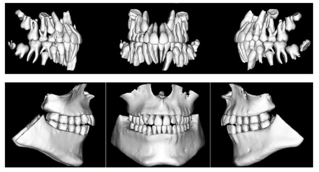
  3. The patient hands your new patient coordinator a disk. The disk contains several digital files.  Among the files are several JPEGs (digital intra and extra oral photographs), two STL files (3D digital study models with centric occlusion bite registration) and one DICOM file (a large field of view CBCT scan).

What happens next depends on your level of familiarity with digital technologies and which orthodontist’s diagnostic records were presented by the patient.  Another question is whether or not the three different sets of diagnostic records satisfy the AAO’s Clinical Practice Guidelines for Pretreatment Unaltered Diagnostic Records – a full PDF of the Guidelines can be found on the AAO Member Website (Clinical Practice Guidelines).   It is my opinion that in order to best serve our patients, we work to establish guidelines to bridge the analog-digital / 2D – 3D divide and create a standard for maintaining, sharing and transferring both analog and digital diagnostic records.

Where do we start?  Analog diagnostic records (plaster study models / radiographic films) seem fairly straightforward: maintain/provide the originals or a satisfactory copy.  Digital diagnostic records are a different story – 2D or 3D – different manufacturers and different software may format the data files into formats that are incompatible with each other.  For guidance, I turned to the medical community, where digital imaging has been commonplace for decades (Digital Radiography, CT Scans, MRI).  A review of the literature (Exchanging and Sharing Medical Images) provided two important considerations: the imaging files should be the original unaltered files and should be provided in a vendor-neutral format.  We didn’t learn about this in our residency programs!

What’s next?  We already discussed analog diagnostic records.  Here’s my opinion on digital diagnostic records. I feel the original unaltered image files should be handled as follows:

  • Intra / Extra Oral Digital Photographs: Should be provided in a JPEG ( .JPG ) file format.  This format is ubiquitous.  You can view this format on your phone or your office workstations.  All imaging software programs will accept photographs in this format.
  • 2D Digital Radiographs: Should be handled the same as digital photographs: JPEG format.  All imaging software and practice management software should have the capability of exporting and importing digital radiographs in JPEG format.
  • 3D Digital Radiographs (CBCT Scans): The original unaltered DICOM ( .DCM ) files should be provided.  The scan data may be exported as a single .DCM file or as a sequence of multiple (hundreds) .DCM files.  All of the major CBCT scanner manufacturers package their scanners with software that can import / export scan data in this format.  My thoughts on accommodations for a non-3D orthodontist’s use of the DICOM dataset will follow.
  • 3D Digital Study Models: The original unaltered intra-oral scan should be provided in STL ( .STL ) file format.  Typically, the Digital Study Models will be exported from the software as two files that contain the upper and lower models plus a bite registration.  Most major 3D intra-oral scanners contain in their accompanying software the ability to convert and export the raw scan data as an STL (vendor-neutral) file set.  If you own a 3D intraoral scanner that has “closed architecture” and cannot export the scan data in STL format, it is my opinion that you should provide a 3D printed set of study models if asked.  The requirements for diagnostic models in the AAO Clinical Practice Guidelines are clear:  they must be 3 dimensional to suffice.  In my opinion, a printed screenshot of digital study models does not meet these requirements.  There are other considerations in handling digital study models (i.e. open shell/surface scan versus closed shell); however, I feel these are beyond the scope of the current discussion.

Now we must consider how all orthodontists, from non-digital to fully 3D digital, can utilize each other’s diagnostic records.  Let’s look back at our scenarios:

  1. The diagnostic records presented in scenario #1 above do appear to fulfill the requirements in the Clinical Practice Guidelines, except that I would like to see the photographs provided in digital JPEG format as opposed to a printout. If the photographs are indeed “photographs,” I have no advice – except to grab your digital camera, cheek retractors and mirrors.  If you receive these records and feel additional information or imaging is required to provide an accurate diagnosis and treatment plan – that is your (and the patient’s) choice.
  2. In my opinion, the diagnostic records presented in scenario #2 do not fulfill the requirements. Diagnostic models need to be three dimensional – a printout is not.  Regarding the other printed pictures – see my recommendations above.
  3. The disc presented in scenario #3 does, in my opinion, fulfill the requirements. The problem is that a non-3D orthodontist is likely unable to utilize or interpret most of the diagnostic information provided.  How then do we bridge this divide?

In my office where we utilize 3D intra-oral scanning and ultra-low dose CBCT scans, the diagnostic records process is fairly quick and relatively non-invasive.  I’ve yet to have a patient say “I’d rather have an impression” after a 3D intra-oral scan.  I have previously blogged about my opinions on ultra-low dose CBCT scanning (AAO Tech Blog).  I understand that not all orthodontists can utilize, or interpret, the data we produce.  When asked to provide a copy of a patient’s diagnostic records, we produce a disc that contains the following files:

  • Intra and extra oral photographs in JPEG format
  • 3D Diagnostic models – original unaltered – in STL format
  • CBCT Scan – original unaltered – in DICOM ( .DCM ) format: this allows another orthodontist to import the scan data into their own 3D viewing software
  • CBCT Scan – with viewer – provided as a separate proprietary file type: this allows another orthodontist (without their own 3D viewing software) to view the scan data
  • Panoramic view – reformatted as a JPEG file
  • Lateral Cephalogram – reformatted as a JPEG file – with distance ruler for tracing purposes

In my opinion, the disc we produce fulfills the requirements for “Pretreatment Unaltered Diagnostic Records,” and as provided, can be utilized by any orthodontist for diagnostic and treatment planning purposes – with one exception:  the STL files.  If physical study models are requested, the STL files should be printed and provided.  Keep in mind most jurisdictions allow for a nominal copying or duplication fee to be assessed to a patient who requests a copy of their records – the 3D printing charges should fall into this category.

The Committee on Technology will be presenting recommendations to the AAO Board of Trustees in the coming months to aid in amending the Clinical Practice Guidelines in regards to Diagnostic Records, Record Keeping and Transfer of Orthodontic Patients.  My thoughts will be a part of the conversation.  It is my opinion that one day, in the not too distant future, our diagnostic records will be all digital and all in 3D.  As this transition continues, I feel we must take steps to bridge this 2D – 3D / analog-digital divide in a manner that is in the best interest of our patients and within the skillset of every orthodontist.

I ask for your comments and suggestions on this topic.  CTech wants to make sure that all AAO Members are represented in the process as our specialty moves forward with advances in technology.


Sharing Cone-Beam CT Images Online

By Dr. Dan Grauer

When diagnosing and treatment planning interdisciplinary patients, have you ever sent your three-dimensional images to a colleague? Have any of your patients requested a copy of their records for a second opinion? Or maybe, a patient declines a radiograph because another orthodontist has recently taken a CBCT image of the patient? In all of these instances, you will need to communicate with the other office to initiate the transfer of CBCT images. The purpose of this blog is to describe different methods used to share patients’ CBCT records via online means.

Images acquired in your office are requested by a second orthodontist/dentist:

The first question that will need to be answered is whether the other office has the possibility of viewing and analyzing the images in three-dimensions. In a few instances, I have found myself trying to transfer a full three-dimensional file, when the second orthodontist just wanted a cephalogram and a panoramic radiograph. If this is the case, your software will probably allow you to create a synthetic cephalogram and panoramic radiograph that can be emailed through a HIPAA-compliant email account. If the second orthodontist requires a three-dimensional image, two case scenarios are possible:

Case scenario 1: Second orthodontist owns software to read and visualize CBCT images.

In this case, your software is able to export the CBCT Images in DICOM format (Digital Imaging and Communication in Medicine). DICOM files are large, and a file transfer application is needed. Once transferred, these can be imported into the software of the second orthodontist for visualization and analysis.

Case scenario 2: Second orthodontist does not own three-dimensional imaging software.

Under this case scenario, the second orthodontist would need both the CBCT images and a three-dimensional viewer. Three main options are available.

Option 1: If you own a CBCT machine, your software is generally able to create a file that includes both the image data and a basic viewer. The files created are large and can be transferred with a file transfer application.

Option 2: Anatomage offers the possibility of uploading your CBCT images to the cloud, and these can be accessed online through Anatomage’s application, which acts as a visualization tool. At this point the software is in Beta-version and can be accessed at www.anatomagcloud.com. You, as the generating office, will need to upload the images to the AnatomageCloud database and use this application to allow the second office to access the specific patient images. The access is granted with a link embedded in an email. After receiving authorization to access the images, the second office will be able to access the images online without the need of downloading them or installing any software.

Option 3: Dolphin Imaging software offers a complimentary viewer, https://www.dolphinusers.com/dolphin-imaging-viewer/. The receiving doctor can view 3D images by downloading and installing the Dolphin Imaging Viewer software. Files are transferred in DAZ file format. This file format is proprietary to Dolphin Imaging, and the files are created by the originating doctor through Dolphin Imaging 3D Software. This option 3 would work also in Case Scenario 1, when both doctors use Dolphin Imaging 3D software, but it is important to note that only the unprocessed images need to be transferred, such as the DICOM file; the viewer is part of the software downloaded by the receiving office.

Images acquired by other offices:

Images that you receive from other offices should be requested in DICOM format. This will permit you to be able to import these into your 3D software. If you obtain the file in a different format than DICOM (that often includes the viewer), the analysis and measurement possibilities are limited; this is because your 3D software most likely includes all the features that you may need while visualizing and measuring 3D Images. If both offices use Dolphin Imaging 3D Software, a proprietary format DAZ can be used to transfer and share images. The advantage of this approach is that all patient images, including both 3D and 2D images, are shared simultaneously.

In summary, with Cone Beam CT becoming more popular in practices, sharing 3D images with other treating doctors or practices requires some additional steps. The first step is to initiate the conversation with the second office to establish the best system to use to share images. The advantages of 3D images over traditional 2D images are beyond the scope of this blog, but once you become accustomed to a transfer and visualization system, the collaboration between doctors and patient care may improve.

Boy, Did I Just Dodge a Bullet!

By Dr. Greg Jorgensen

(Although you may be more tech-savvy than my mom, you may still work with an older doctor, team member, or relative who could benefit from this story.)

I was seated in church when I got the call from my 80-year-old mother. She said, “Boy, did I just dodge a bullet!”

When I asked her what happened, she explained that she was surfing the web looking for a recipe when a “loud warning” popped up on her screen. It said something like, “Your computer has been infected with a virus that could destroy your files. Call Microsoft support immediately at 1-222-333-4444 (not the actual number).”

She said that the message was accompanied by a “blaring siren” that scared her and that no matter what she tried, she could not close the alert popup. Worried about losing her files, she quickly called the “support line” number on the screen.

The voice on the other end of the line identified himself as a “Level 7” Microsoft technician. He told her not to worry, that her files were safe and that he would solve her problem. She dutifully followed his instructions which allowed him to remotely connect to her computer. She was so proud that she was able to follow his instructions without my help. After several minutes of watching windows open and close on her screen, he told her that everything was now working and her files were safe. He also notified her that she didn’t have antivirus software installed and that for a one-time fee of $699 she could purchase lifetime support and protection from future attacks. Not wanting to go through this again, she read him her Discover Card account number over the phone.

Upon hearing her story, I immediately had her disconnect her computer from the Internet and shut it down. I explained that she had been scammed and that everything the “technician” told her was a lie. It took me over an hour to convince her that he was not legitimate. Once I had her laptop disconnected from her WiFi, I started damage control. I tried to log on to her retirement account website and found that it had already been locked down because of multiple attempts to log on using the wrong password. I found the same to be true for her bank account. I assume that the scammer had obtained login information from her browser history.

We immediately called all of her financial institutions and credit card companies to lock down her accounts. We were even able to get Discover to stop payment on the “support fee” she paid. My mom didn’t lose any money, but the inconveniences she’ll face over the next month or two will be a big hassle.

When I asked her why she didn’t call one of my siblings or me like she usually does when she has computer issues, she said that the siren scared her and that she trusts Microsoft (even though she was browsing the Internet on her MacBook and wasn’t even using Microsoft software!) She also thought she was safe because she placed the phone call rather than received it, even though she got the number from the fake alert. In review, my mom made three glaring mistakes – 1) she called an unverified number, 2) she helped them log onto her computer, and 3) she gave them her credit card number.

This “phishing” technique is just one of many being used by scammers to steal from less-than-tech-savvy people. The appropriate way to get out of this situation would have been to simply choose “Force Quit…” under the Apple Menu or right click on the taskbar in Windows and choose “Task Manager.” These options would have allowed my mom to close the browser along with the ominous alert popup. Had she done this, the scammer would not have even known she saw the alert message.

A quick review of safe surfing practices with your team or relatives should include the following recommendations:

  1. never click on a link that arrives in an email unless you are 100% sure that it is legitimate and you are expecting it (don’t just trust the name on the email…scammers can hack your friends’ email accounts).
  2. never open an attachment that arrives in an email unless you are 100% sure of its origin and are expecting it.
  3. never call a phone number supplied in an email or alert box (instead, look up the support number yourself if you feel you need to call). Be leery of any email that arrives unexpectedly or doesn’t look or feel right. If you aren’t certain, don’t click, open, or call.

Big Data Revisited

By Anthony M. Puntillo DDS, MSD

In August 2014, I wrote an introductory article for this blog entitled “What is Big Data and How is it Related to the Practice of Orthodontics?” As more orthodontic practices move to the digital collection of orthodontic treatment records (EHRs-photos, models, radiographs, treatment history) and more of our data is being stored in the “cloud”, there is a tremendous opportunity for us as a profession to access that data for the betterment of our patients and advancement of our specialty. Over the last ~3.5 years, however, there has been little visible traction by our researchers and leadership on this front. Meanwhile, there should be no doubt that corporate entities (DSOs and orthodontic vendors) understand the value of our data. Check out the recent cover article for Fortune Magazine (“Tech’s Next Big Wave: Big Data Meets Biology” -3/19/2018). The article notes that “The quest to retrieve, analyze, and leverage (medical) data has become the new gold rush.” If orthodontists are to hope to have any influence on how orthodontic treatment is delivered in the future, management of our patients’ data will be crucial. Technology has sped up every aspect of our lives. We must now start to give this issue the attention it desperately demands. But where should we begin?
If we are to tackle this challenge, there are many complex questions that will need to be answered. Our patients’ privacy is not the least of these. Even with our busy professional and personal lives, I imagine it has been hard for most to miss the recent public flogging of Mark Zuckerburg and Facebook. Both he and his company were taken to task by Congress and the media when they revealed that the personal data for 87 million of their customers had been inappropriately accessed by an outside research company. As a result of these disclosures, politicians are threatening regulations for their industry – think HIPAA for Silicon Valley. There are more than 2.5 million people who annually seek orthodontic treatment. As we look to find the best ways to utilize our patient’s treatment data to improve their care, we must make certain that it is being done in a way that is respectful of all patients’ privacy. While privacy may be where we start, there are other, even more difficult issues that need to be addressed.

The complexity of the Big Data issue will require the input from the brightest minds both from within and from outside of our profession. To that end, the Great Lakes Association of Orthodontists has put forth a resolution to this year’s American Association of Orthodontists House of Delegates. Resolution 18-18 GLAO (http://hod.live.aaoinfo.org/resolution2/18-18-glao-big-data-task-force-and-records-repository/) requests that our Association President appoint a Big Data task force. I would encourage you to review the resolution and let your representatives (HOD Delegates) know your feelings. While this issue is certain to require a significant investment of time, talent and financial resources, we cannot afford to leave this investment to outside sources. Those who control the data will control the future.

Is your Password Already Floating Around on the Internet?

By Steve McEvoy, Technology Consultant

Passwords are a pain. You need them when you turn your computer on, open your practice management software, access your email and when you access most any other Internet service like Gmail, Pandora, Dropbox, Facebook, etc. Keeping track of all of them is a hassle, and it is human nature to look for shortcuts – many people often use the same password for multiple sites.

Hackers are constantly looking for ways to steal information. Information is the new ‘gold’ on the Internet. The mention of ‘hacker’ conjures up images of a mysterious character lurking in a dark room, presumably hard at work trying to guess your username and password to gain access to your information. While this may still be the case in some situations, the hackers are smart folks, and they have moved on to where the real gold is. Rather than hacking us one person at a time, they are going after the websites where all of our collective online information lies.

Do you have an online account with any of these sites? LinkedIn, Yahoo, Dropbox, Adobe, Target, Home Depot, Comcast, Bell, Equifax or Experian? What all these sites (and many others I haven’t listed) have in common is that they were hacked, and some of the valuable information stolen included your username and passwords for their site. Ten’s of millions of usernames and passwords have been stolen. The hackers have realized that putting their efforts into breaking into a website yields much more information about you than trying to hack you directly.

How do you know if your username and password was breached?

Can you rely on the hacked website to notify you? Some sites, when they discover they have been hacked, implement a mandatory password change the next time you attempt to access the site. Has this ever happened to you? You log in to a website, and it immediately prompts you to verify your identity and change your password? It did for me a while back when I was using Dropbox. What they didn’t point out was that they had been breached, and for some period of time hackers could have accessed my data.

Can you even rely on the websites to know when they have been hacked? How would they know? It’s not like a traditional crime where you might see the broken window. Companies that aren’t making security a principal focus may be completely unaware of the breach and your user information for that site might be already out in the wild.

Troy Hunt is a security expert at Microsoft, he’s one of the white hat hackers on our side. He had the great idea to compile a list of all the available hacked accounts he could find. He scoured the ‘dark web’ to get copies of the information being sold by successful hacks (there is a thriving retail market for this fueled by BitCoin). He found nearly 5 billion accounts (that’s a B, not an M) from 265 known breaches. Then he created the website “Have I been Pwned” (www.haveibeenpwned.com). That’s not a typo, “pwned” is a slang online gaming term that roughly means “I own you” or “I conquered you” just like a hacker may have. His website is free to all. You can go to the site, enter the username that you may often use online (for example most people use their email address) and it will tell you if it knows your username was leaked in one of the breaches it knows about. I tried it with mine and found my information was leaked in the Adobe and Dropbox breaches.

You can also enter a password to see if the password is already in the known hacked password list. In the example shown here, I am testing the password that Invisalign Intraoral scanners use by default. Pwned.

A word of caution. Should we really trust that whoever is behind the website isn’t recording all the passwords tried? What if they get hacked? My advice is to be careful here and NOT test any of your CURRENT passwords you use where you have precious information kept (like your online bank account password). I know this is counter-intuitive, this is the first password you want to test to see if it’s safe.

Remember the bad habit that people have using the same username and password at multiple sites? If that’s you, and the hackers have got your username along with the password when they hacked one of these sites, I can guarantee you that these are the first things they are trying at other websites to see if they can get in (perhaps your bank). If the usernames and passwords are the same, they get immediate access without even needing to take a second guess. It happens all the time.

Consider all this carefully. Check the email you use typically for a user account at HaveIBeenPwnd.com. Perhaps check a password you use all the time. If you discover you have been pwn’d, change your passwords at all the sites that share that username immediately.

Facebook as an Education/Networking Tool

By Katie Graber, DDS, MS

Traditionally orthodontic professionals have relied upon refereed journals, scientific lectures or national/regional orthodontic meetings to garner information about the latest and best in treatment. In the last decade, many orthodontists have transitioned to attending distance-based seminars, listening to podcast recordings or following online group discussions in their area of interest. This, coupled with an increased attendance at specific vendor meetings, has changed the way information is communicated in our field to a portion of the membership.

One of the newer “remote” vehicles for information transfer is via private or closed Facebook groups. These groups can have a narrow or large focus. Some are grouped regionally, by alma mater, treatment style, etc. They can range in focus from practice design to technology to specific treatment products and/or mechanics.

Some forums that I follow include:

  • Elevate Orthodontics Podcast (1700+ members)
  • Ortho Cosmos Forum (1200+ members)
  • Jeff Nolte: Suresmile (300+members)
  • Women in Orthodontics (1200+ members)
  • AlignerInsider: Clinical Chat (1300+members)

Others include:

  • OrthoPreneurs (1600+ members)
  • The Pragmatic Orthodontist: Clinical (3000+ members)
  • The Pragmatic Orthodontist: Elite (150+ members)
  • Orthodontic Pearls (1800+ members)
  • OrthoX Elite
  • Ortho Aligner 3D Study Group
  • Ortho Voice
  • Sarver Orthodontic Study Group
  • Suresmile Synergy
  • Trios Orthodontic Study Group

These groups provide orthodontic professionals the opportunity to network online through a free source. Facebook closed groups permit the owner to determine the eligibility of the membership as well. Vetting members can take time and many of the host/owners do this on a voluntary basis. Additionally, the forum allows for easy photos, video and recording attachment and is an “in real time” communication tool for peer-peer discussions.

These orthodontic peer groups can get information to the user immediately. There is no waiting for a national or vendor meeting. While mostly anecdotal in nature, the “real life” experience of peers is a valuable source for cutting-edge advances. Sometimes the groups also use collective bargaining and/or group purchasing to earn discounts on products or services relevant to that group. Some I have seen include Mari’s list, 3D printers (Form2) and do-it-yourself aligner software. The discounts are above and beyond what a single orthodontic practice can earn or negotiate as the power comes from the sheer number of people interested/included in the discount group. This is another potential benefit to joining one of these online groups.

The Orthodontic Podcast group allows members to listen to orthodontists discuss current and relevant topics. This is reminiscent of the AAO Practical Reviews audio-cassette recordings of years past; albeit these have no continuing education credit associated nor are endorsed/reviewed/refereed by the AAO. A benefit of this type podcast (versus waiting for the practical reviews publication) is instant listening, the instant ability to download and listen across a broad spectrum of listening devices/locations. In addition, there is generally no fee (per se) to join the Facebook group nor listen in on many of the podcast recordings.

So, if you love “talking teeth” and have a specific interest; there is probably a Facebook group for you! Search your subject area under “groups” on Facebook and ask to join.


CBCT Imaging for your Practice: Is Now the Time?

By Dr. Kenneth R  Webb

As I walked the exhibit floor at the AAO Annual Session in San Diego this past April, I stopped and talked to several of the company reps displaying the latest generation of CBCT scanners. Competition is good – what struck me is how far this technology has come in such a short period of time. Effective dose, image quality, and versatility are three areas where the advances are most impressive. I have witnessed these advancements first hand in my practice where we have been using CBCT since 2012 and recently upgraded a satellite office from a 2D digital Pan / Ceph to an “Ultra Low Dose” current generation CBCT scanner. I’d like to share some of my thoughts about the 2D to 3D transition.

Effective Dose
I encourage anyone interested in CBCT technology to search for studies authored by Dr. John Ludlow and his team. Dosimetry data and image quality for many CBCT scanners (both older and current generations) have been researched in great detail. Comparative data for 2D dental radiographic imaging (both intra and extra oral) is also available. By replacing our 2D Digital Pan / Ceph with a current generation CBCT scanner we have reduced the effective dose of our diagnostic records imaging by approximately 60%. (Ludlow JB, Walker C. AJO-DO, 2013;144 (6): 802-817) This is accomplished with one, approximately 5 second, ultra-low dose CBCT scan compared to the two longer duration exposures (pan then ceph) required with 2D imaging. Certainly a win-win for our patients.

Versatility
What else can we use the 3D data for? Digital models: used to assess the inter-arch and intra-arch relationship as well as aiding the determination of arch length and arch width requirements and assessing arch symmetry. Modelling labs can produce articulated 3D study models from the CBCT data (DICOM) files.

STL files of the patient’s dentition and occlusion can be uploaded into whatever software you may be using in your office for 3D model storage and viewing. Additionally, these models can be used (with appropriate software) to produce treatment simulations by “moving” individual teeth or the arches as a whole. Imagine completing your diagnostic records with Intra / Extra Oral photos and one 5-second CBCT scan!

Viewing the Diagnostic Data
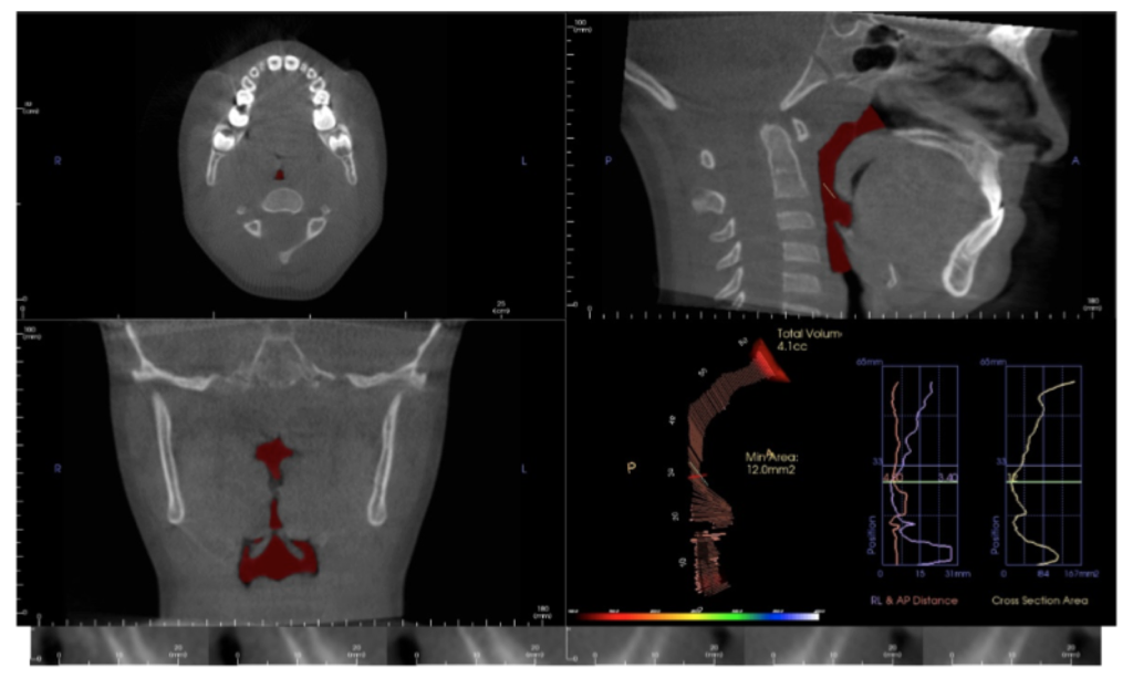
The software that comes with a CBCT scanner can easily produce the customary 2D diagnostic images that we are used to evaluating (panoramic, lateral and A-P ceph). In addition – you see everything! The data can be viewed in sagittal, axial or coronal sections (slices) or as a 3D “volume rendering” which can be rotated and sliced (“clipped”) at will. So why is this additional information gained from CBCT imaging important?

Pathology
You will see pathology in the 3D data that isn’t visible with standard 2D imaging. When pathology is visible in 2D, the 3D data can more accurately ascertain location, extent, and character of the area of concern. This is beneficial to our patients.
Developmental Anomalies
One of my first revelations when we began imaging in 3D was the prevalence and extent of “individual anatomic variation”. But how much is too much? We have made referrals to medical specialists for significant developmental anomalies in the cervical spine, nasal cavity and paranasal sinuses. Patients / parents are appreciative of the thoroughness of our diagnostic process that includes 3D imaging.

Asymmetries
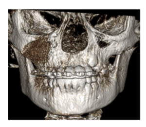
They get their own paragraph! Your patient smiles at you and you notice a vertical asymmetry and occlusal plane “smile” cant. Is it caused by hemimandibular hyperplasia, condylar hyperplasia, a unilateral expansive lesion in the maxilla, a growth response to unilateral progressive condylar resorption (to name a few) or a growth response to a foreign object lodged in the nasal cavity? I’ve seen them all. Similar clinical presentations – different treatment plans.

TMJ
Condylar position in centric occlusion, condylar size and shape, glenoid fossa morphology, condylar osseous morphology, joint space, findings suggestive of degenerative joint disease – both active and stable. It is hard to argue that these are not important considerations in our diagnostic process.

Airway
The value of volumetric and cross-sectional analysis of the naso – pharyngeal airway and its relationship to orthodontic diagnosis and treatment planning has, and continues to be, studied in great detail. Is the growth and development of a patient who presents with narrow arches, an anterior open bite or anterior crossbite, tongue thrust and a strong mouth breathing pattern secondary to adenoid / tonsillar hyperplasia, a deviated nasal septum, environmental allergies or restrictive airway dimensions in general? Should your imaging modality include an assessment of these areas?

And there is more…
Orthognathic surgical treatment planning, TAD placement guidance, precise localization of ectopic and supernumary teeth, and assessment of treatment progress – including evaluation of root torque. All are possible or enhanced with 3D imaging. And by managing scan parameters (field of view, scan time and voxel size) at an effective dose equal to or less than 2D imaging modalities.

So, if you haven’t brought this technology into your practice – is now the time?

At the 2017 AAO Winter Conference in Ft. Lauderdale, Mr. Chris Bentson reported on a survey of recent (2016) U.S. Orthodontic Residency graduates: 88% responded that they had used CBCT imaging for diagnosis and treatment planning during their residencies including 21% that used it on all patients.

For the 2013 graduates, the responses were 76% and 9% for the same questions.

Based on these statistics, are we that far away from 3D imaging being included in the “standard of care” discussion? Embracing new technology is not easy but the value added for the practitioner and our patients is significant. The orthodontic educators who expertly taught my generation faced a similar situation during their careers with a then relatively new technology: panoramic radiographic imaging. My generation faced the challenges of transitioning from analog imaging (film) to digital.

Change is not easy. The transition from 2D to 3D imaging in your practice will require a financial and educational commitment. The whole staff will be a part of this paradigm shift. A positive – 3D imaging will be a differentiator for your practice. If you are seeking an associate, partner or complete transition, 3D imaging will make your practice more attractive to the next generation of orthodontists.

3D imaging will benefit you and your patients. Is now the time?

What Your Email Address Says About You

By Steve McEvoy, Technology Consultant

Sending business emails ending with @Netcom.com, Aol.com, Earthlink.com and soon Yahoo.com are tell-tale signs you aren’t keeping up. People notice and may ask you if you are still driving the ‘72 Ford Pinto as well.

Using personal accounts from your Internet Service Providers (ISP) also looks old school like Comcast.net, TimeWarner.com, RoadRunner.com, SBCGlobal.net.

There is no rule making us keep up, or against driving a ‘72 Pinto, but I’d argue if you are in a marketing battle for new patients, this is just one small aspect you can easily improve.

For work, you really should have an email for your practice’s domain.   [email protected] or [email protected], for example. People expect this, and when they see [email protected], you send a subtle message that isn’t positive.

You might certainly have a private, personal email account as well, but even for that, you should consider using something like [email protected] or [email protected].

This should also apply to any of your staff using email on behalf of the practice.

There may be other motivating reasons as well. Companies offering email services for free (AOL and YAHOO) that aren’t thriving financially in their core business won’t be putting development resources into keeping products current, safe and secure.  The news is full of stories about hacked email accounts and ransomware which typically makes its way into your computer via email. Companies like Google, Microsoft, and Apple have reputations to maintain and will have the resources to keep up.  If you are going to use a free email service, I might suggest you look to one of the major players.

Changing your primary email account is always a major hassle, and this is likely what has kept you from making the switch.  Setting up a new email account, and merely forwarding all the email from your old account indefinitely isn’t the right solution. Your old email account can still be hacked, and the company can still go out of business.

The steps generally are:

  • Setup your new email account – and take this opportunity to make sure the password is a hard one. I’d suggest you setup two-factor authentication with it as well.
  • Link your new account to your PC, phones, tablet, etc.
  • Using your old email, notify EVERYONE in your contact list that you are changing your email to the new one effective immediately.  You can do this in one mass email, but be smart about it and put all the recipients in the BCC list so they don’t see everyone else you emailed the list to. Make yourself the only official To:
  • Configure your old email account to forward to your new one (for a while).
  • After a month, check your old account to see if anyone you know is continuing to use the old account, then contact them directly to start using the new address.
  • After another month, turn off the forwarding from your old account and delete it at the vendors. Gone forever, but no risk of hacking.

You can also always enlist some help from your IT person, they should be well familiar with the process.

Creating Videos for Your Orthodontic Practice

Humans are visual creatures. Simply put, we process visual data better. Research shows that more than 80 percent of human learning is done visually and that humans process visuals 60,000 times faster than text.1  As such, creating compelling, easily sharable videos is one of the best ways you can choose to promote and showcase your orthodontic practice. Thankfully, creating low cost, high impact videos online has never been easier. With just a small amount of effort and know-how, you can make a lasting, positive impression on potential patients and set your practice apart from competition in your area.

Videos that introduce yourself and your staff, feature patient testimonials and showcase your practice and the services you offer is a key way to build trust and let potential patients get to know you before actually ever stepping foot in your office. As a bonus, videos hosted on your practice website can also improve how your site appears in search engine results…meaning increased web traffic.

Videos can easily be created without a major investment in equipment or software.  Although they do take some planning and time, especially early on, the investment is well worth the effort. But, if you are trying to keep your costs low, a newer model iPhone or Android device you may currently have in your pocket offers outstanding video capabilities. (Just be sure you’re shooting in horizontal vs. vertical mode.)

Regardless of what equipment you use, following these basics.

  • Shooting a video can be intimidating at first. Just always try to focus on sound and picture quality.
  • Shoot your video in 720p or 1080p resolution.
  • Pre-plan your video. Have a goal in mind. Create and work from an outline so you don’t go off-track.
  • Be sure to add captions to your video. This is because up to 85 percent of videos watched and shared socially are viewed without.  Captions are added via a video editor’s text feature.
  • Make the first few seconds of your video visually impactful. Your goal is to capture your viewer’s attention quickly, within the first 3 seconds.
  • Give your video a compelling title or headline.
  • Brevity is key. Be efficient with words and visuals.

As you begin planning your videos, consider:

  • Stability is key. Invest in a small tripod, monopod or desk stand – your videos will be of much higher quality if you can prevent excess camera movement.
  • If you are producing a video for patient education, you may want to invest in a high quality consumer camcorder with an external microphone.
  • While professional lighting is always preferred for marketing and practice website videos, natural lighting is usually sufficient. Just film in a brightly lit room and make sure there is enough light on your subject’s face.
  • Make sure to have your interviewee or one of their parents sign a photo/video release form giving you permission to use the video you just shot. These forms are available at aaoinfo.org. Log on; then go to Practice Management > Forms and Releases. Scroll down to Practice Management Forms & Materials; then click on Photo Releases.
  • Think outside the box. Videos can be an extremely powerful communication tool for patient education and staff training.

Once you’ve captured your video, you’ll want to begin editing. Editing video means trimming and arranging your video. Video editing software is also how you can add transitions, music, captions and titles…as well as export the movie file that you’ll host on your website or social media channel. Thankfully, there are easy-to-use and affordable options available that will allow you to quickly transform your raw footage into a finalized video for your web page or social media channels.

For users on the Mac, iMovie is a solid choice for beginners. On the Windows side, Windows Movie Maker is an ideal tool to use. Some video users even use apps on their smart phones to edit footage. Apps like the iMovie app for the iPhone and CyberLink PowerDirector for Android are well worth considering for no-frills, simple video edits. For more advanced editing, you might consider options like Apple’s Final Cut Pro X or Adobe’s Premiere Pro CC.

Creation of videos goes quickly and the learning curve is shallow. Make it a goal to begin producing videos today. Fight the urge to think it’s too difficult and just get started.

REFERENCE
1. Gutierrez, Karla. “Studies Confirm the Power of Visuals in eLearning.”

Shift eLearning (8 July 2014). Retrieved from shiftelearning.com/blog/.
* For advice or encouragement on how you can easily begin shooting videos for your practice, or for any video related questions you have, please contact Burt Bollinger, AAO Multimedia Production Manager at [email protected].